
Videos
To review:
The sequence of events that occurs during the cardiac cycle through flow chart and the indication of contraction of auricle and ventricle and their relaxation.
Introduction:
Circulatory system circulates the blood into two directions simultaneously, one from the heart to the body known as systematic circulation and the other from the body to the heart, from where it goes to the lungs for purification called the pulmonary circulation and the whole process is known as double circulation.
Explanation:
The right side of the heart contains blood which is poor in oxygen and the left side contains oxygen-rich blood, the atrioventricular (AV) valve and semilunar valves that are present between the auricle and ventricle on their side and at the opening of pulmonary artery and aorta, respectively. These prevent the back flow in the heart. The right auricle is usually filled with the deoxygenated blood by superior or inferior vena cava and the left auricle is filled by the oxygenated blood coming from lungs by the pulmonary veins.
The contraction (systole) of auricles on both sides start after they are completely filled with the blood and the blood enters in their corresponding ventricles through AV valves, as soon as the ventricles are filled up and starts contracting. The AV valves are closed to prevent the back flow of blood and starts auricular relaxation. The blood from right ventricle moves to the lungs for its purification through pulmonary artery and oxygenated blood from left ventricle is transported to other body parts through the aorta. This completes one cycle of double circulation of blood in the body and it is known as a cardiac cycle, and this cycle is continuous. The diagram below shows the steps of cardiac cycle:
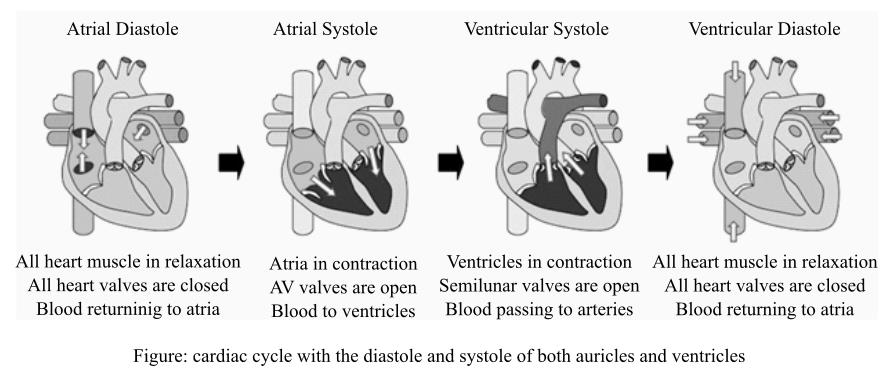
Diastole means relaxation of the auricles and ventricles and systole is the contraction of auricles and ventricles. Atrial diastole occurs when the blood returns to the atria from vena cava on right side and from pulmonary vein on left side. The valves of the heart, during this time, are closed and muscles are relaxed. When the atria are filled, the AV valves open and atrial systole occurs. The ventricular systole occurs when they are completely filled and semilunar valves open and blood releases out from the ventricles and cause diastole of ventricles. This way the cardiac cycle goes on till the heart is functional.
Explanation:
The right side of the heart contains blood which is poor in oxygen and the left side contains oxygen-rich blood, the atrioventricular (AV) valve and semilunar valves that are present between the auricle and ventricle on their side and at the opening of pulmonary artery and aorta, respectively. These prevent the back flow in the heart. The right auricle is usually filled with the deoxygenated blood by superior or inferior vena cava and the left auricle is filled by the oxygenated blood coming from lungs by the pulmonary veins.
The contraction (systole) of auricles on both sides start after they are completely filled with the blood and the blood enters in their corresponding ventricles through AV valves, as soon as the ventricles are filled up and starts contracting. The AV valves are closed to prevent the back flow of blood and starts auricular relaxation. The blood from right ventricle moves to the lungs for its purification through pulmonary artery and oxygenated blood from left ventricle is transported to other body parts through the aorta. This completes one cycle of double circulation of blood in the body and it is known as a cardiac cycle, and this cycle is continuous. The diagram below shows the steps of cardiac cycle:
Diastole means relaxation of the auricles and ventricles and systole is the contraction of auricles and ventricles. Atrial diastole occurs when the blood returns to the atria from vena cava on right side and from pulmonary vein on left side. The valves of the heart, during this time, are closed and muscles are relaxed. When the atria are filled, the AV valves open and atrial systole occurs. The ventricular systole occurs when they are completely filled and semilunar valves open and blood releases out from the ventricles and cause diastole of ventricles. This way the cardiac cycle goes on till the heart is functional.
Want to see the full answer?
Check out a sample textbook solution
Chapter 13 Solutions
Human Physiology


- Label the hearts main parts in the diagram below.arrow_forwardExplain the electrical and mechanical events in the cardiac cycle, including their relationship to the electrocardiogramarrow_forwardIn the figure below on the left, label the P, QRS and T waves. Describe what is happening in the heart in the P wave: Relate the P wave to the cardiac cycle:o Is the heart in systole or diastole?o Is the pressure high or low?o Where is blood flowing? Which valves are open? closed? o Which muscle fibers are contracting, if any?arrow_forward

 Human Physiology: From Cells to Systems (MindTap ...BiologyISBN:9781285866932Author:Lauralee SherwoodPublisher:Cengage Learning
 Fundamentals of Sectional Anatomy: An Imaging App...BiologyISBN:9781133960867Author:Denise L. LazoPublisher:Cengage Learning
- Basic Clinical Lab Competencies for Respiratory C...NursingISBN:9781285244662Author:WhitePublisher:Cengage
 Human Biology (MindTap Course List)BiologyISBN:9781305112100Author:Cecie Starr, Beverly McMillanPublisher:Cengage Learning